Unmasking the Illusion of Brain-Like AI: York University Study Reveals Critical Flaws in Visual Neural Networks

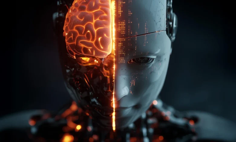
For over a decade, the intersection of neuroscience and artificial intelligence has been defined by a compelling and transformative narrative: that artificial neural networks (ANNs), engineered to mimic human vision, are the closest digital approximations of the biological brain. These algorithms, which excel at object recognition, image classification, and pattern extraction, have become foundational tools for neuroscientists seeking to decode the mysteries of human perception. However, a groundbreaking study conducted by researchers at York University challenges this core assumption, revealing a profound and previously overlooked asymmetry in how artificial intelligence and biological brains process visual information.
The research, published in the prestigious journal Nature Machine Intelligence, demonstrates that while AI models can successfully predict brain activity, the reverse process does not hold true. Recorded neuronal responses cannot reliably predict the internal features of these AI models. This revelation exposes a critical vulnerability in modern computational neuroscience: an AI system can yield output that mimics biological behavior while relying on entirely alien internal mechanisms. As researchers increasingly turn to machine learning to model not only vision but also language, hearing, and motor control, this discovery forces a fundamental reassessment of how artificial intelligence is validated and utilized in scientific inquiry.
The Paradigm Shift: Testing AI From the Opposite Direction
To understand the weight of the York University findings, one must examine the methodological history of computational neuroscience over the past ten years. Traditionally, neuroscientists investigating visual processing have operated within a unidirectional framework. Researchers record the electrical activity of neurons—typically in the visual cortex of primates—while subjects are exposed to various stimuli. They then train artificial neural networks on similar visual tasks and test whether the mathematical activations within the layers of the AI can predict the recorded biological brain activity.
For years, high correlation scores in these forward-prediction tests were interpreted as proof of architectural and operational equivalence. If an ANN could successfully forecast how a neuron would fire in response to an image of a car or a bear, scientists assumed the model was utilizing algorithms structurally or functionally akin to those of the primate brain.
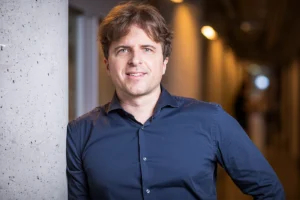
Led by Assistant Professor Kohitij Kar, the York University research team decided to test the robustness of this assumption by inverting the investigative lens. Kar, who serves as the Canada Research Chair in Visual Neuroscience and holds appointments at York’s Centre for Vision Research and Centre for Integrative and Applied Neuroscience, collaborated with postdoctoral fellow Sabine Muzellec—a trainee within the university’s Connected Minds program—to ask a reverse question: Can recorded biological brain activity accurately predict the internal features and states of the artificial intelligence models?
If these computational systems truly operate like the biological brain, the researchers reasoned, the relationship should be mutually predictive. To evaluate this hypothesis, the team developed an innovative diagnostic framework termed reverse predictivity testing. This methodology was engineered to uncover hidden discrepancies that conventional, one-way alignment tests systematically overlook.
Rigorous Testing Across Diverse Visual Stimuli
To conduct their evaluation, the York University team exposed both biological subjects and artificial neural networks to a meticulously curated dataset of visual stimuli. The testing suite comprised 1,320 natural photographs and realistic synthetic images depicting ten familiar categories: bears, elephants, human faces, apples, cars, dogs, chairs, airplanes, birds, and zebras. These objects were placed against a wide array of complex natural backgrounds, as well as controlled indoor and outdoor environments.
To further challenge the systems, the researchers introduced 300 additional images modifying the same subjects across various artistic and simplified visual styles. These included stark outlines, technical drawings, schematic representations, and abstract artistic interpretations. By evaluating how both biological brains and ANNs processed these variations, the team could map the boundaries where computational mimicry began to diverge from biological reality.
The results of these trials were striking. While the artificial intelligence models continued to demonstrate a strong capacity to predict the activity of biological neurons—replicating the success metrics that have validated ANNs in literature for years—the inverse operation failed significantly. Recorded neuronal responses proved markedly less effective at predicting the internal computational features of the models.
Crucially, this failure was unique to the comparison between biology and machine learning. When the researchers cross-compared neuronal activity recorded from one biological brain against that of another biological brain, no such predictive asymmetry appeared. Brains could reliably predict other brains, but brains could not reliably predict the inner workings of AI models that ostensibly simulate them.
Underlying Mechanisms and Scientific Implications
The implications of this unidirectional predictive failure are profound. The findings indicate that contemporary artificial intelligence systems are successfully solving visual recognition tasks by employing internal strategies and computational shortcuts that the biological brain does not utilize.
"The results were striking. While AI models can predict the neurons we recorded in the brain fairly well, the brain cannot equally predict many of the model’s internal features. Interestingly, this is not the case when neurons from one brain are compared against ones from another brain," explains Kar.
This divergence suggests that surface-level behavioral matching—such as correctly identifying an object in an image—is an insufficient metric for validating cognitive models. An AI can arrive at the correct answer through a flawed, non-biological pathway. As artificial intelligence models grow exponentially more complex, relying solely on forward-prediction metrics risks compounding these hidden errors. Over time, as researchers continuously refine ANNs based strictly on their ability to mimic output rather than underlying process, the gap between artificial models and actual primate brains will only widen.
This widening chasm directly threatens one of the primary justifications for integrating machine learning into neuroscience: the desire to construct empirically accurate, mechanistically valid models of the human nervous system. If the internal mechanics of the AI do not reflect biological reality, using them to theorize about human cognition risks building theoretical frameworks on faulty foundations.
Broader Impacts on Clinical Research and Human Behavior
The fallout from this study extends far beyond theoretical computer vision and neurophysiology. In recent years, artificial intelligence has woven itself into the fabric of clinical research, behavioral psychology, and neurological diagnostics. AI models are routinely deployed to help design experimental paradigms, simulate human behavioral responses, and investigate complex psychological and neurological conditions, ranging from neurodevelopmental disorders to trauma-related conditions.
Many of these sophisticated applications rest on the foundational premise that the underlying code processes information in a manner structurally akin to human cognitive architecture. If that premise is proven false, the scientific community faces the risk of drawing erroneous conclusions from models that exhibit high surface-level accuracy while operating on fundamentally different computational principles.
This concern is particularly acute in sensitive clinical domains, such as autism research. Accurate computational baselines of neurotypical brain function are vital for researchers attempting to isolate how perception, sensory processing, and information integration differ in neurodivergent populations. If the baseline model is driven by non-biological AI strategies, the resulting clinical insights could be skewed.
"Our findings challenge how similar current AI systems really are with the primate brain. We show that models that were previously thought to be brain-like rely on internal components that the brain does not appear to use. We provide a well-vetted diagnostic metric for the field," notes Muzellec.
Furthermore, these cautionary findings are not isolated to the visual cortex. Similar artificial neural network architectures are currently deployed to study auditory processing, linguistic comprehension, and motor control. If those specialized systems similarly rely on internal computations divergent from biological neural pathways, the neuroscience community may need to reevaluate the extent to which current machine learning paradigms can genuinely illuminate the nervous system.
A Path Forward: Building Better Models Through Diagnostic Tools
Importantly, the York University study does not advocate for the abandonment of artificial neural networks in neuroscience. Rather, it provides the academic and development communities with a much-needed diagnostic toolkit designed to bridge the gap between machine learning performance and biological fidelity.
By deploying reverse predictivity testing, researchers can systematically isolate which specific layers, nodes, and features of an artificial neural network genuinely correspond to authentic neural activity, and which are artifacts of non-biological optimization. This granular mapping allows computational scientists to filter out misleading models and concentrate engineering efforts on developing architectures that more faithfully reflect biological perception.
"Our approach helps identify which parts of an ANN truly match brain activity, allowing us to build more reliable models for understanding how people see and interpret the world," states Kar. "This is especially important for our autism research program, which builds on models of the neurotypical brain as a baseline."
To accelerate this corrective process and ensure the broader scientific community can scrutinize their own models, the York University research team has made their evaluation methodology publicly accessible. Developers and neuroscientists can now utilize a dedicated testing toolkit released via PyPI to audit their AI models, measure reverse predictivity, and identify targets for architectural improvement.
Ultimately, the study published in Nature Machine Intelligence marks a maturing of the relationship between artificial intelligence and neuroscience. It signals a transition away from uncritical acceptance of AI as an electronic replica of the mind, and toward a rigorous, diagnostic-driven approach. By demanding that artificial intelligence not only reaches the correct perceptual conclusions, but does so through mechanisms that genuinely mirror human biology, researchers can pave the way for a new generation of computational models that advance both computer science and our understanding of the human brain.