Why Ancient Vision Systems Shaped the Brains Crossed Wiring Across Half a Billion Years of Evolution

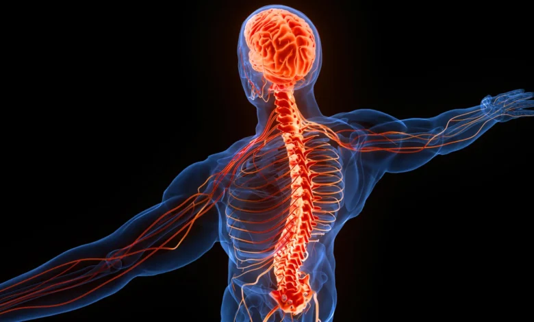
For centuries, anatomists, physicians, and evolutionary biologists have pondered one of the most persistent structural puzzles of human and vertebrate biology: why the left cerebral hemisphere controls the right side of the anatomy, while the right hemisphere governs the left. This contralateral organization—where sensory inputs and motor commands cross the body’s midline—has long eluded a definitive functional consensus. While previous theories have suggested that crossed wiring might optimize computational efficiency, prevent neural bottlenecks, or aid in spatial navigation, a new study offers a compelling alternative rooted in deep evolutionary history. Researchers propose that this unusual neurological arrangement is not necessarily an optimized adaptation for survival, but rather an architectural inheritance dating back more than 500 million years, anchored to the emergence of primitive vertebrate visual systems.
The investigation, spearheaded by Dr. Martin Wessendorf, a neuroscientist and associate professor emeritus at the University of Minnesota known for his work on spinal cord neuronal regulation, alongside Daniel Wang, a specialist in cognitive neuroscience and neurodegenerative disorders, was recently published in the peer-reviewed journal Frontiers in Neuroanatomy. By examining multiple neural pathways across tetrapods and other vertebrate lineages, the research team mapped how modern central nervous systems may have expanded outward from an ancient visual blueprint. Their findings suggest that as evolutionary pressures added complex sensory and motor circuits to early organisms, these new pathways conformed to the pre-existing spatial maps established by ancient visual processing centers.
Main Facts and the Mechanics of Contralateral Wiring
To understand the scope of the researchers’ hypothesis, one must first examine the physiological mechanisms of contralateral processing in living vertebrates. The phenomenon begins at the point of sensory input. When photons pass through the lens of an eye, the resulting projection onto the retina is inverted. Environmental stimuli originating on an organism’s right side register on the left hemiretina, whereas stimuli on the left project onto the right hemiretina.
This inverted spatial map is preserved as signals travel along the optic nerve to the optic chiasm, an anatomical junction where nerve fibers intersect or continue uncrossed depending on the species. Through this arrangement, visual data from the right visual field is directed to the left hemisphere of the brain, and vice versa. Beyond the optic chiasm, these signals project into a midbrain structure known in mammals as the superior colliculus, and in non-mammalian vertebrates as the optic tectum. As one of the most evolutionarily conserved structures in the vertebrate brain, the optic tectum is responsible for generating immediate, reflexive orienting movements—such as instantly turning the head toward a sudden flash of light or an unexpected shadow in the periphery.
Crucially, the optic tectum does not process sight in isolation. Deeper strata within the tectum integrate auditory, tactile, and proprioceptive inputs, organizing these diverse sensory modalities into coherent spatial maps that align directly with the visual representation of the opposite environment. The researchers noted that this multi-sensory integration hub set a precedent for how incoming data and outgoing motor commands were spatially segregated across hemispheres.
A Timeline Spanning the Cambrian Period
The chronological framework of this evolutionary hypothesis reaches far back into the Earth’s history, targeting the Cambrian Period roughly 538 to 486 million years ago. During this epoch of rapid biological diversification known as the Cambrian explosion, the earliest vertebrate body plans began to take shape among primitive, jawless aquatic organisms.
According to the study’s authors, the widespread presence of an optic chiasm and a functionally organized optic tectum across every living vertebrate lineage implies that these foundational structures must have been present in the common ancestors of modern fish, amphibians, reptiles, birds, and mammals. Because these structures predated the evolution of terrestrial life, limbs, and complex cerebral cortices, they established a structural template long before higher-order cognitive processing emerged.
As tetrapods—four-limbed animals—transitioned from aquatic environments to land approximately 370 to 350 million years ago, their nervous systems faced new mechanical and sensory demands. Yet, rather than engineering entirely novel architectures from scratch, evolution appears to have retained the foundational crossed layout established by ancestral visual systems. Subsequent sensory and motor tracts, including the dorsal column pathway, the spinothalamic tract, the tectospinal pathway, and the rubrospinal pathway, all adopted this contralateral configuration.
Tracing Sensory and Motor Pathways Across Species
The research team systematically analyzed the structural anatomy of major sensory and motor tracts to determine how deeply conserved these crossed pathways truly are. Their findings highlight a clear dichotomy in how different systems cross the central nervous system midline.
Sensory Pathways:
The dorsal column pathway, which transmits fine touch, conscious proprioception, and vibration, travels upward through the dorsal region of the spinal cord before synapsing in the brainstem and crossing to the contralateral hemisphere. While absent in fish, this pathway is reliably identified in amphibians, reptiles, birds, and mammals, indicating that it emerged early in the tetrapod lineage and has remained remarkably stable. Conversely, the spinothalamic tract—responsible for conveying pain, thermal sensations, and crude touch—exhibits an even deeper evolutionary lineage. It crosses the midline directly within the spinal cord itself and is found not only in tetrapods but also in certain cartilaginous fish, such as sharks and rays, pointing to origins that predate the vertebrate land invasion.
Motor and Movement Pathways:
Motor control exhibits a similarly complex web of crossed connections. The tectospinal pathway, which facilitates rapid behavioral responses to unexpected environmental stimuli (such as evading a sudden predator), routes movement signals from the deep layers of the optic tectum across the midline to the opposite side of the spinal cord. Similarly, the rubrospinal pathway, originating in the midbrain red nucleus, extends contralaterally to assist in limb coordination, appearing prominently in mammals and select other tetrapods and cartilaginous fish.
The Evolutionary Trajectory of the Corticospinal Tract
Perhaps the most intricate evolutionary narrative uncovered by the researchers involves the corticospinal tract, the primary neural highway responsible for voluntary motor control, and notably, the fine, dexterous manipulation of human fingers.
While all vertebrates possess a telencephalon—the anterior region of the brain that forms the basis of the cerebrum—only mammals feature a neocortex characterized by six distinct cellular layers. This six-layered architecture enabled advanced cognitive functions, including sensory-memory integration, abstract planning, and language.
In most non-primate mammals, the corticospinal tract primarily serves a regulatory function, projecting to sensory relay nuclei in the spinal cord to modulate incoming tactile information. However, in primates—and most notably in humans—this pathway expanded its operational scope to form direct monosynaptic connections with motor neurons controlling the distal musculature of the hands and digits.
The Minnesota researchers hypothesize that rather than inventing a completely novel motor circuit to accommodate complex tool use and precise manipulation, human evolution repurposed an existing, ancient neural pathway. By adapting a circuit initially optimized for sensory regulation or generalized movement, evolution endowed the corticospinal tract with a secondary, highly specialized function.
Expert Analysis and Broader Implications
The publication of this hypothesis has drawn commentary from broader neuroscientific and evolutionary biology communities, prompting a re-evaluation of teleological assumptions in biology. Historically, evolutionary adaptation has frequently been viewed through a purely utilitarian lens—the assumption that every anatomical quirk must confer a direct, measurable survival advantage.
However, Dr. Wessendorf and Wang’s findings underscore the profound power of evolutionary conservatism and path dependency. The researchers argue that the brain’s crossed wiring may not be superior to an uncrossed system in terms of computational speed or physical utility. Instead, it represents a historical contingency: once the ancient visual system established a crossed organizational scheme, subsequent neural circuits followed suit simply because building upon an existing template was genetically and developmentally more parsimonious than designing a new architecture.
From an analytical perspective, this hypothesis offers testable parameters for paleoneuroscientists and evolutionary anatomists. The authors suggest that future research could focus on examining the fossilized neurocraniums and endocranial casts of ancient jawless fish and stem-gnathostomes from Cambrian and Ordovician fossil beds. Discoveries of preserved soft tissues or structural impressions denoting the presence of an optic chiasm and midbrain visual maps would provide empirical support for the timeline proposed in the study.
Furthermore, understanding the evolutionary scaffolding of the central nervous system has broader implications for clinical neurology and neurorehabilitation. As researchers continue to map the developmental genetics that guide axonal pathfinding and midline crossing during embryonic growth—such as the roles played by netrins, slits, and other molecular guidance cues—recognizing that these pathways are beholden to ancient architectural constraints may help illuminate why certain neural injuries or congenital malformations manifest the way they do.
Ultimately, the study bridges the gap between microscopic neuroanatomy and deep evolutionary history. It reframes the human brain not as a flawlessly engineered machine built from a blank slate, but as a deeply layered palimpsest. Every time a human reaches out a right hand to grasp an object, controlled by the left hemisphere of the brain, that action is a silent echo of visual processing systems that guided ancient aquatic organisms through primordial oceans more than half a billion years ago.