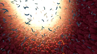
Decoding the Enemy Within: Johns Hopkins Researchers Uncover How a Common Gut Bacterium Drives Colon Cancer and Develop a Promising Molecular Shield

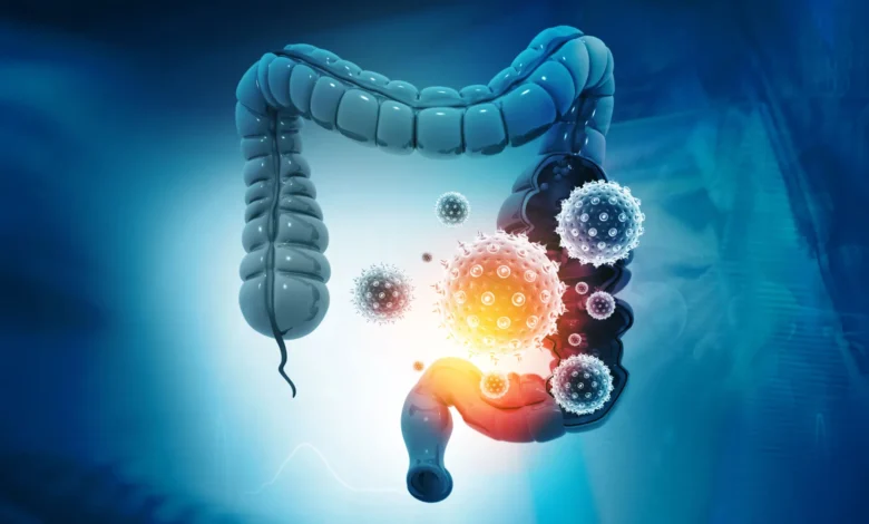
For nearly two decades, the scientific community has harbored a quiet and complex concern regarding a microscopic resident of the human digestive tract. Bacteroides fragilis, a bacterium that comfortably colonizes the intestines of up to 20% of healthy individuals, possesses a dark side. While it often exists harmlessly as part of a diverse microbiome, certain strains secrete a potent agent known as B. fragilis toxin, or BFT. For years, researchers have known that this specific toxin damages the delicate lining of the colon, triggers chronic inflammation, and actively drives the formation of malignant tumors. Yet, despite extensive investigative efforts, a foundational mystery persisted: how does the toxin initially anchor itself to intestinal cells to launch its devastating assault?
That persistent gap in medical knowledge has finally been bridged. In a breakthrough published in the journal Nature, an interdisciplinary team of researchers led by scientists at the Johns Hopkins Kimmel Cancer Center, the Bloomberg~Kimmel Institute for Cancer Immunotherapy, and the Johns Hopkins University School of Medicine has identified the missing link. The team discovered a specific protein, claudin-4, that acts as the essential docking port for BFT. More remarkably, the researchers have already leveraged this discovery to engineer a molecular decoy that intercepts the toxin before it can compromise intestinal tissue, successfully protecting animal models from harm and charting a potential new course for colorectal cancer prevention.
The Chronology of a Decades-Long Investigation
To appreciate the magnitude of this recent breakthrough, it is necessary to examine the timeline of scientific discovery that paved the way for it. The journey began in earnest during the late 2000s, an era when researchers were increasingly recognizing the profound influence of the human microbiome on systemic health and disease.
In a landmark 2009 study published in Nature Medicine, Dr. Cynthia Sears and her colleagues at Johns Hopkins provided some of the first definitive evidence that BFT could actively promote the development of colon tumors. This pivotal work established a concrete mechanistic link between toxin-producing gut bacteria, sustained localized inflammation, and the initiation of colorectal cancer—a malignancy that remains one of the leading causes of cancer-related mortality worldwide.
Subsequent investigations during the 2010s focused on the downstream effects of the toxin. Scientists discovered that BFT systematically dismantled E-cadherin, a critical structural protein responsible for holding neighboring intestinal cells together and preserving the integrity of the colonic epithelial barrier. When E-cadherin is cleaved and destroyed, the tight barrier fails, allowing luminal bacteria, microbial byproducts, and inflammatory molecules to infiltrate underlying tissues.
However, a frustrating paradox baffled investigators. Although BFT clearly targeted E-cadherin, biochemical analyses revealed that the toxin did not bind directly to it. Researchers hypothesized that an intermediary molecule—a distinct receptor on the cell surface—must be capturing the toxin and facilitating its attack. Repeated attempts over many years to isolate and identify this elusive receptor repeatedly fell short, creating a major bottleneck in the development of targeted preventative therapies.
Leveraging CRISPR to Solve a Biological Mystery
The breakthrough finally materialized when the Johns Hopkins team, collaborating with researchers at Harvard Medical School, harnessed the unprecedented precision of CRISPR gene-editing technology.
Maxwell White, an M.D./Ph.D. candidate in the Sears laboratory, spearheaded a genome-wide CRISPR screen alongside the laboratory of Dr. Matthew Waldor at Harvard. By systematically deactivating individual genes within colon epithelial cells—the frontline cells that form the inner lining of the large intestine—the researchers sought to determine which specific genetic instructions were absolutely required for BFT to attach and inflict damage.
Out of thousands of candidates, one gene emerged as a resounding and unambiguous top hit: the gene responsible for producing claudin-4. In healthy cellular architecture, claudin-4 is a foundational building block of tight junctions, specialized membrane structures that seal the spaces between adjacent cells and regulate the passage of ions and molecules through the tissue barrier.
When the researchers experimentally disabled the claudin-4 gene, the dynamic shifted entirely. BFT lost its ability to bind to the colon cells. Deprived of this essential initial anchorage point, the toxin was completely rendered incapable of reaching or cleaving its downstream target, E-cadherin. The mystery of the missing receptor was solved: claudin-4 was the mandatory gatekeeper through which the bacterial toxin initiated its destructive cycle.
Challenging Dogmas: An Unexpected Cellular Mechanism
The identification of claudin-4 as the BFT receptor caught the research team by surprise, defying established biochemical expectations.
Prior to the screen, scientists widely anticipated that BFT would interact with a classical cell-signaling protein, such as a G protein-coupled receptor. These signaling receptors typically act as molecular communication switches, transducing extracellular cues into internal cellular responses. Claudin-4, by contrast, is primarily a structural protein designed to maintain physical adhesion and barrier integrity between cells, rather than mediating complex biochemical signaling cascades.
Furthermore, the mechanism highlights a rare operational strategy for bacterial toxins. BFT belongs to a class of enzymes known as proteases, which function by cutting other proteins. The vast majority of proteases interact directly with their intended targets. BFT, however, utilizes an unconventional two-step approach: it must first dock onto a completely separate structural receptor (claudin-4) before it can successfully execute its proteolytic strike on E-cadherin. A comprehensive review of existing microbiological literature revealed no other known bacterial toxin utilizing this precise mechanism, underscoring the novelty of the discovery.
To confirm the physical interaction between the two molecules with absolute certainty, the Johns Hopkins team partnered with structural biologists F. Xavier Gomis-Rüth and Ulrich Eckhard at the Molecular Biology Institute of Barcelona. Through advanced biochemical assays designed to measure molecular binding affinities, the researchers demonstrated that BFT and claudin-4 form a tightly integrated complex in a strict one-to-one stoichiometric ratio, providing definitive physical proof of their affinity.
Translating Discovery into Defense: The Molecular Decoy
In medical research, identifying a disease mechanism is valuable; translating that mechanism into a tangible defense is transformative. Armed with the knowledge that BFT relies exclusively on claudin-4 for attachment, the Johns Hopkins team conceptualized a strategy to trick the toxin.
If the bacterium’s weapon requires claudin-4 to dock, what would happen if the colon was flooded with a decoy version of that same protein?
To test this hypothesis, the investigators engineered a soluble molecular decoy featuring specific sequences derived from claudin-4. This engineered protein was designed to mimic the natural receptor, floating freely within the intestinal environment to intercept BFT before it could reach the actual cellular lining.
Partnering with the laboratory of Dr. Min Dong at Harvard Medical School—including researcher Kang Wang and colleagues—the team tested the decoy strategy in murine (mouse) models. These in vivo experiments allowed scientists to observe the toxin’s behavior within the complex biological ecosystem of a living mammalian intestine.
The results exceeded expectations. When introduced into the system, BFT bypassed the natural claudin-4 receptors on the colon epithelial cells and instead bound avidly to the circulating decoy proteins. By successfully intercepting and neutralizing the toxin in transit, the decoys shielded the mice from BFT-induced tissue damage and barrier degradation.
Official Responses and the Path Forward
Reflecting on the milestone, senior author Dr. Cynthia Sears emphasized the broader significance of the findings for human health.
"We’ve made several attempts over time to identify the receptor, so this is an exciting moment," says Dr. Sears, who serves as the Bloomberg~Kimmel Professor of Cancer Immunotherapy and professor of medicine at Johns Hopkins. "Understanding how bacterial toxins work can open doors to new approaches for detection and therapy for associated diseases, including diarrhea, colorectal cancer and bloodstream infections."
Maxwell White, the study’s lead author, highlighted the translational potential of the team’s decoy strategy, noting that the current iteration represents just the starting point for pharmacological development.
"This approach could be iterated upon with small molecules or other biologics that have better pharmacological properties," White explains. The research team is currently evaluating various molecular formats to determine which pharmacological structures will optimize the stability, delivery, and efficacy of the decoy in clinical settings. Small-molecule compounds and engineered biologic proteins are both under active consideration as researchers work to optimize the therapy for potential human application.
Broader Implications for Oncology and Infectious Disease
Despite the enthusiasm surrounding the discovery, the scientific team maintains a measured and rigorous perspective regarding the road ahead. Important technical hurdles and transitional phases remain before this strategy can influence human medicine.
While the mouse models conclusively demonstrated that the molecular decoy protects against acute tissue damage, the current studies did not explicitly measure the long-term prevention of colorectal cancer. Bridging that gap will require extended longitudinal studies tracking tumor incidence in animal models exposed to toxigenic Bacteroides fragilis.
Additionally, structural biologists have yet to capture the exact, high-resolution three-dimensional structure of the BFT-claudin-4 complex. Even state-of-the-art computational tools, such as AlphaFold, have thus far been unable to fully resolve the precise atomic geometry of how the two molecules interlock. Gaining this structural clarity could empower researchers to use structure-based drug design to engineer even more potent and specific inhibitory compounds.
Nevertheless, this breakthrough resolves a fundamental biological question that has persisted in oncology and microbiology laboratories for nearly two decades. By mapping the exact point of attachment utilized by a ubiquitous gut pathogen to disrupt human tissue, researchers have transformed an abstract microbial threat into a tangible, targetable vulnerability.
As investigations continue and funding from institutions such as the National Institutes of Health, the Bloomberg~Kimmel Institute for Cancer Immunotherapy, Cancer Research UK, Janssen Research and Development, and the Howard Hughes Medical Institute continues to support these collaborative efforts, medicine moves one step closer to intercepting cancer-promoting pathogens before their destructive cascade can ever begin.