Unlocking Nature’s Ultimate Fiber: How Scientists Decoded the Atomic Secret of Spider Silk

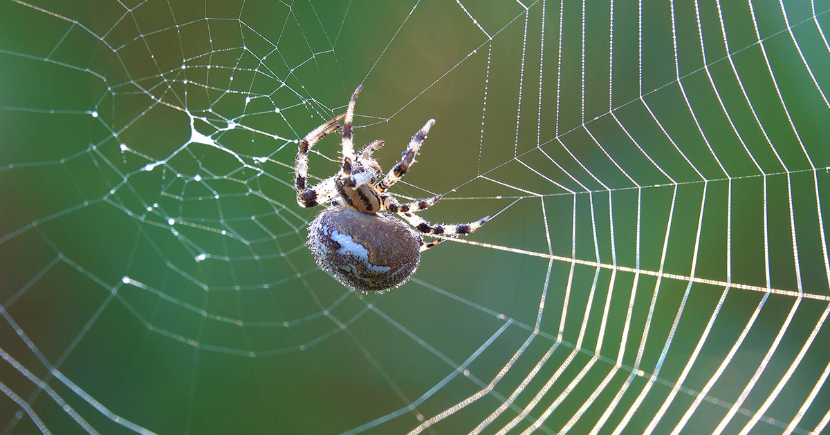
When a spider plummets from a wall, disaster is averted in a fraction of a millisecond. With a flick of its spinnerets, the arachnid spins a microscopic soup of proteins into climbing gear that is tougher than Kevlar and stronger than structural steel. For decades, materials scientists have viewed spider silk as a biological holy grail—a material boasting exceptional tensile strength alongside high flexibility and biocompatibility with the human body. Yet, despite decades of study, duplicating this marvel in a laboratory setting has remained an elusive goal.
The primary hurdle has been a fundamental gap in scientific understanding: researchers simply did not know how a spider transforms "silk dope"—a liquid blend of proteins, water, and salt held within its silk glands—into a remarkably resilient solid fiber upon extrusion.
Now, a team of chemists and spectroscopists at San Diego State University has bridged that divide. By probing the molecular structure of native silk at the atomic level, the researchers have uncovered how a chaotic tangle of unstructured proteins orchestrates a phase transition into a strong, flexible fiber. The findings illuminate a widespread biological strategy used across diverse organisms and offer a crucial roadmap for manufacturing next-generation bio-inspired materials.
Inside the Spider’s Silk Factory
Spiders are nature’s most versatile micro-manufacturers, capable of producing up to seven distinct types of silk depending on the species and immediate environmental needs. Aciniform silk is deployed to wrap and immobilize prey; stretchy flagelliform silk reinforces the shock-absorbing spirals of capture webs; and rigid dragline silk acts as the primary safety line anchoring both the web and the spider to vertical surfaces.
The structural foundation of these fibers was first identified in 1907, when researchers discovered that spider silk is composed of long, modular proteins known as spidroins. These proteins belong to the same overarching family of fibrous structural proteins that includes human collagen and keratin. Decades later, X-ray diffraction studies revealed that the solid silk fiber is built from flat sheets of proteins—termed beta sheets—embedded within a less ordered, non-crystalline matrix.
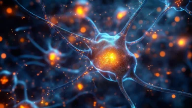
However, a paradox confounded researchers entering the 2000s. When scientists inspected spidroins in liquid solution, they failed to find the rigid, fixed structures characteristic of typical folded proteins. Instead, the proteins featured intrinsically disordered regions that fluttered constantly, dynamically forming and dissolving transient shapes. For years, the mechanism by which this disordered protein soup organized into high-performance solid fibers remained a mystery.
A Chronology of Discovery: From Micelles to Condensates
The quest to understand spider silk formation mirrors a broader paradigm shift in molecular biology over the last two decades. Early theories in the field proposed that liquid dope proteins aligned progressively like liquid crystals, or that they organized into spherical micelles—similar to the proteins found in silkworm silk—which subsequently compressed during extrusion.
A pivotal turning point arrived in 2007, when researchers observing laboratory-engineered spidroins noticed that the proteins spontaneously formed liquid-like globs. Shortly thereafter, in 2009, scientists at the Marine Biological Laboratory in Massachusetts and the Max Planck Institute in Germany identified droplets of aggregated RNA molecules inside fertilized worm eggs. These were recognized as biological condensates: liquid- or gel-like bubbles of concentrated proteins suspended in solution.
Though such structures had been observed by microscopists for over a century, the 2009 discovery heralded a new understanding of condensates as critical organizational tools for cellular life. Biologists soon realized that condensates concentrate molecules, pre-organizing them into malleable microenvironments without locking them rigidly into place. Living cells utilize condensates everywhere—to package DNA, regulate gene expression, and assemble cellular machinery. Macro-organisms also leverage the strategy to build structural wonders, from the stiffness gradients in squid beaks to the rock-anchoring adhesives of mussels and the protective sand-cement tubes of marine worms.
By 2020, biomaterials chemist Keiji Numata of Kyoto University proposed that spider silk undergoes a similar condensate phase during its transformation from liquid dope to solid thread. Numata hypothesized that phosphate ions residing within the ampulla—the middle section of the spider’s silk gland—might trigger this transition via liquid-liquid phase separation. Yet, the precise atomic-scale mechanisms driving the phenomenon remained completely unmapped until recently.
The Black Widow Experiment: Bridging In Vivo and In Vitro
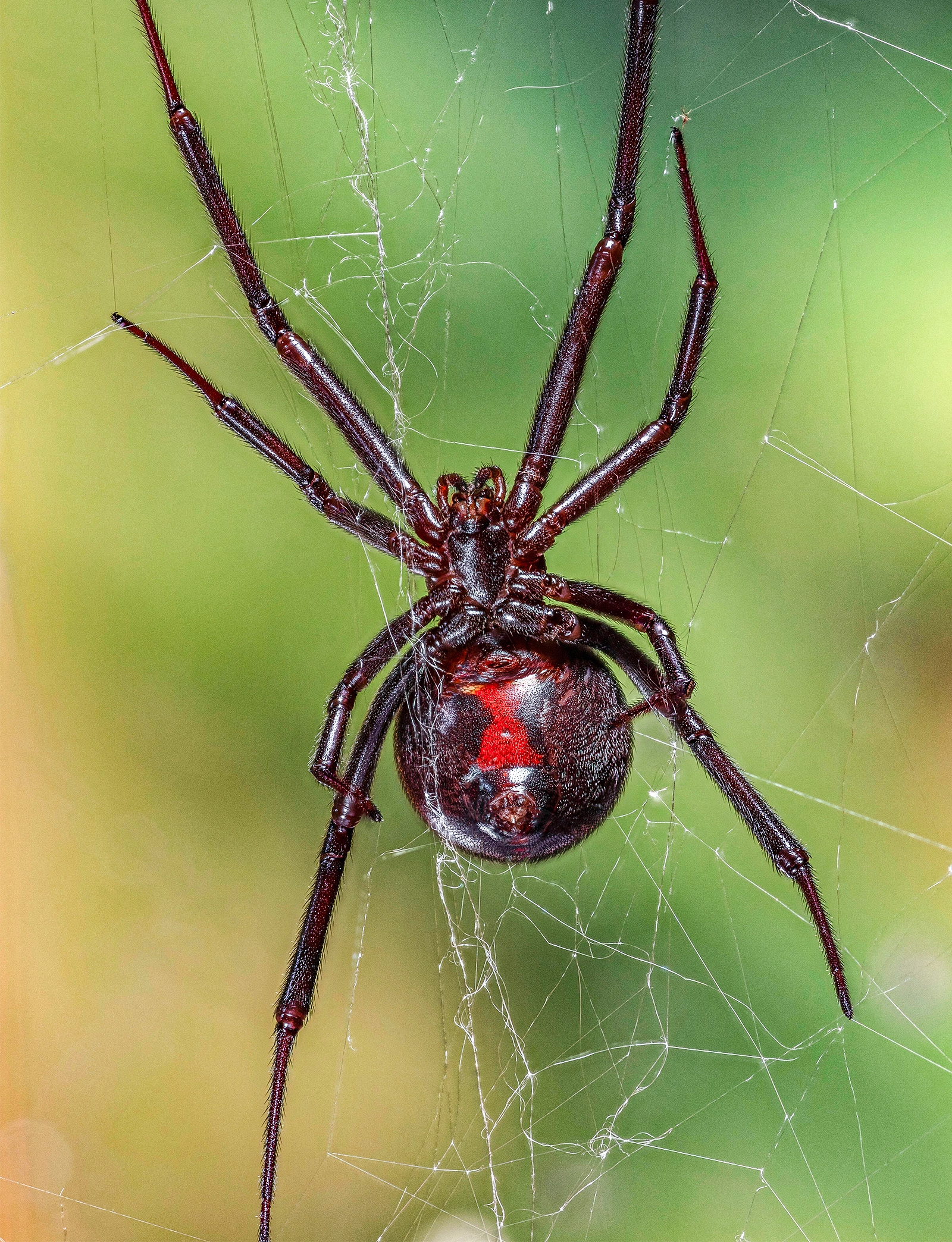
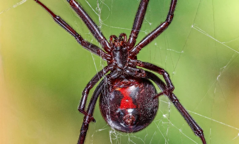
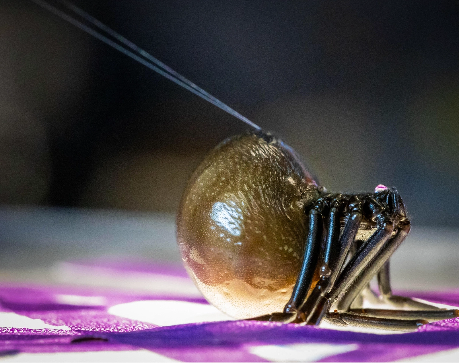
To capture the missing atomic details, a research team led by Gregory Holland at San Diego State University turned to a dependable laboratory subject: the black widow spider (Latrodectus). Famed for their potent venom, black widows are ideal candidates for silk research because their dragline spidroins are among the best-characterized in arachnology, and the spiders can be ethically sourced year-round.
Throughout 2023 and 2024, shipments of female black widows arrived regularly at the university’s chemistry department. Over a series of controlled procedures, graduate student Hannah Johnson and her colleagues gently anesthetized the spiders using carbon dioxide. Wearing protective gear, the researchers used fine tweezers to manually pull a strand of dragline silk from the spider’s spinnerets, reeling it continuously around a motor-driven vial.
During the extraction process, the spiders were maintained on a specialized diet—effectively "protein shakes" enriched with specific amino acid isotopes that could be tracked analytically. Johnson and her team then utilized nuclear magnetic resonance (NMR) spectroscopy to map the molecular structure of the liquid spider dope and the condensates harvested directly from the spiders’ ampullae. Simultaneously, Kevin Chalek, then a postdoctoral researcher in Holland’s lab, mapped the atomic structure of the proteins within the solid, extruded fiber.
"We were trying to bridge the gap between what is happening when the protein is inside the spider and when it’s outside the spider," Chalek explained. "These are the same protein, so how can they be so different? And what exactly is happening between the two?"
Molecular Stickers and the Mechanics of Phase Separation
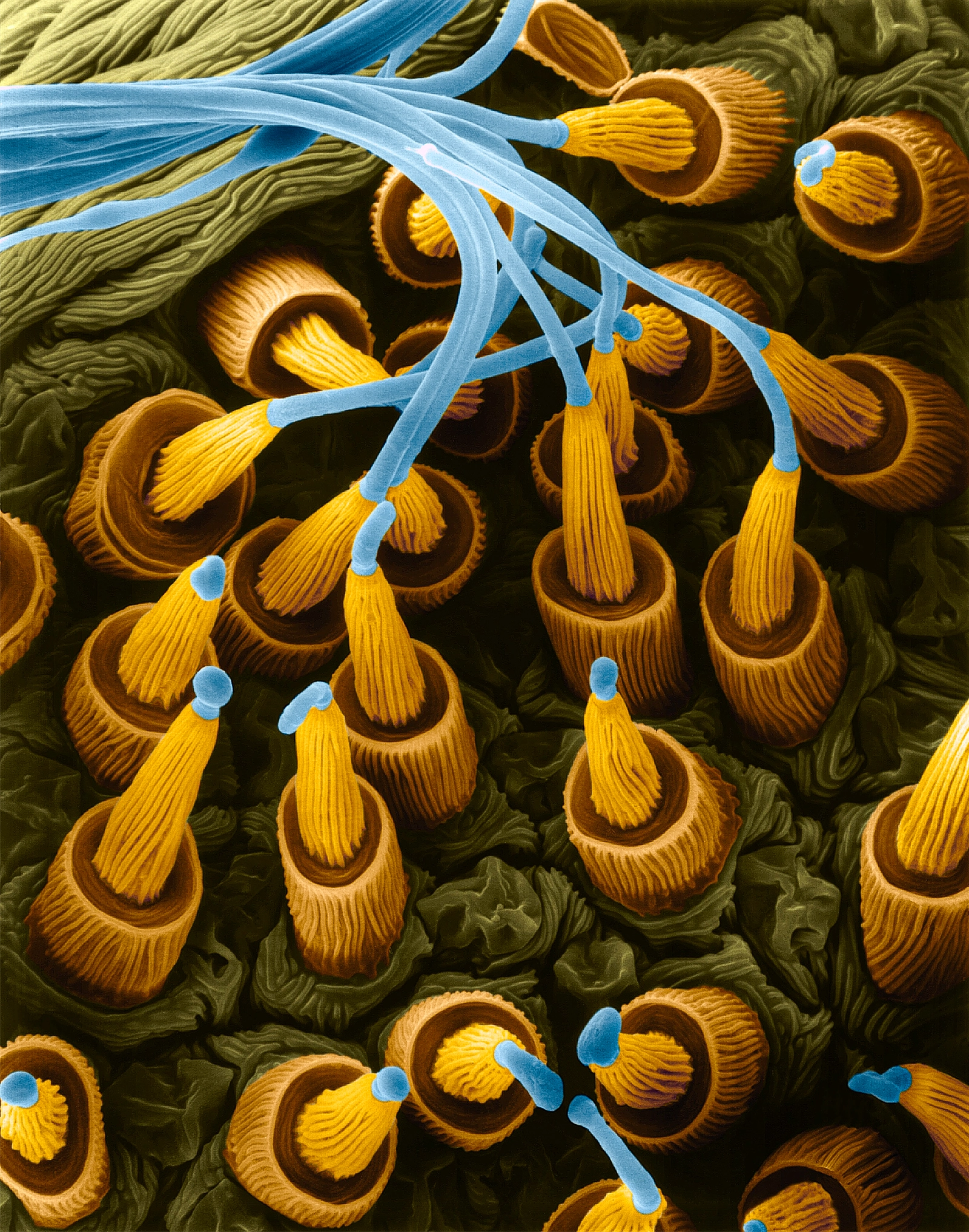
Through high-resolution NMR analysis, Johnson observed that introducing phosphate to the liquid silk dope initiated the condensation process. Specifically, the side groups of two distinct amino acids—arginine and tyrosine—located within the intrinsically disordered regions of the spidroin proteins began to interact weakly with one another.
These weak interactions displaced surrounding water molecules, causing the disordered proteins to pack more closely together into a dense, gel-like condensate. Crucially, these same amino acid interactions persisted even after the material solidified into a finished fiber. This observation strongly implied that arginine and tyrosine residues act as "molecular stickers," guiding the otherwise chaotic proteins first into functional condensates and ultimately into rigid structural fibers.
To validate these findings computationally, other members of the research team deployed advanced artificial intelligence tools, including AlphaFold for protein structure modeling, alongside molecular dynamics simulations. The computational models confirmed that these critical amino acid side groups line up precisely at the bends of the beta sheets in the final fiber, proving their direct role in directing the spidroins into their ultimate architecture.
Independent experts hailed the work as a major milestone. Matthew Harrington, a biological materials scientist at McGill University who was not involved in the study, noted that the research highlights a remarkably common biological strategy. Daiwen Yang, a biologist at the National University of Singapore, described the findings as "direct evidence" that condensates form naturally from native silk, marking a significant step forward for biomaterials science.
Reconciling Competing Theories of Silk Formation
The discovery of condensate involvement does not invalidate earlier models of silk spinning; rather, it refines them. Gregory Holland, who previously demonstrated the presence of micelles in native spider silk, suggests that micelles, condensates, and liquid crystals likely represent consecutive or overlapping stages of a single, highly dynamic phase transition.
In the liquid dope, micelles may prevent messy, disordered proteins from prematurely tangling. As processing continues, proteins pre-assemble into condensates or align into liquid crystal structures immediately prior to extrusion. According to Holland, the actual process inside the living gland is likely heterogeneous and fluid, with multiple structural configurations coexisting simultaneously rather than occurring in a rigid, linear sequence.
"I don’t think these are mutually exclusive," Harrington agreed. "You might have different sorts of phase transitions that are occurring at different stages of the processing."
Despite these breakthroughs, significant questions remain. Researchers have yet to map out every amino acid interaction occurring along the biological assembly line. Furthermore, while phosphate is a known catalyst, scientists suspect additional chemical or physical triggers ("switches") remain undiscovered within the gland’s microenvironment.
Implications for Next-Generation Materials
Translating these biological blueprints into commercial manufacturing could revolutionize materials science. While silkworms have been successfully farmed for millennia to produce commercial textiles, scaling spider production is notoriously difficult. Because spiders are intensely cannibalistic, they cannot be housed communally; farming them requires individual containers, and extracting usable silk yields far lower volumes per animal. Producing a single silk cape, for instance, requires harvesting from roughly one million spiders.
Consequently, the future of spider silk lies in bio-synthetic manufacturing. However, human attempts to replicate spider silk by synthesizing identical amino acid sequences in the lab have historically yielded fibers that fall short of natural performance. Synthetic silks lack the full tensile strength and elasticity found in nature, hinting that crucial structural nuances—or specific minor proteins left out of synthetic formulas—remain missing from current manufacturing recipes.
By decoding the atomic roadmap of how nature uses condensates and molecular stickers to build high-performance materials under ambient conditions in aqueous environments, researchers are drawing closer to replicating the process. Unlike Kevlar and other synthetic high-strength polymers, which demand extreme temperatures, high pressures, and corrosive acidic baths during production, spiders manufacture superior materials cleanly and efficiently at room temperature.
"Once we really understand those blueprints," said Chris Holland, a biologist at the University of Sheffield, "we’ll be able to do more stuff with silk."