
Michigan’s Medicaid Eligibility System Under Scrutiny as IT Glitches Deny Vital Care to Disabled Residents


Marie Noon, a resident of Michigan, navigates a daily regimen of eight crucial medications. These prescriptions are not mere conveniences; they are lifelines, each designed to manage severe health conditions including a heart condition that necessitates the prevention of dangerous rate spikes to avert stroke, debilitating migraines, and the dangerous accumulation of excess fluid in her body. These medications are a direct consequence of adult-onset Still’s disease, a rare and aggressive form of inflammatory arthritis that, over a decade ago, fundamentally altered Noon’s life.
Before her diagnosis, Noon led a vibrant suburban existence in Michigan. Her days were filled with the typical demands of raising two children—shuttling them to cheerleading practice, choir rehearsals, and track meets. She was an active participant in the Parent-Teacher Association and managed a local bank branch. This picture of active community involvement and professional responsibility was shattered by the onset of her illness. The progression of Still’s disease was swift and brutal, reducing her from a capable, active individual to someone who, at the height of her illness, could barely crawl to the bathroom, enduring constant pain and crying uncontrollably from being so sick.
The severity of her condition rendered Noon unable to work for eight years, a period punctuated by prolonged hospital stays that sometimes stretched for weeks. The psychological toll was immense, leading her to confide, "I honestly thought I was going to die."
The shock and disbelief intensified when, after losing her private insurance, Michigan denied her application for Medicaid benefits. The situation was exacerbated by the revelation that this critical denial stemmed from an apparent information technology error, as later clarified by an attorney who assisted Noon in overturning the decision. "I can’t afford my medical care. I have to have insurance," Noon stated, her voice underscored by the urgency of her situation, especially now that she has returned to work.
At the heart of this systemic issue lies the complex and vast Medicaid eligibility system in Michigan, a system that has been managed by Deloitte, a global consulting firm, for nearly two decades. Since 2006, Deloitte has secured contracts totaling approximately $768 million to operate Michigan’s Medicaid eligibility system. This involvement is not confined to a single state; Deloitte plays a dominant role in government IT contracts nationwide, having been awarded contracts in at least 25 states to develop or manage the crucial computer systems that govern access to essential safety net benefits, including Medicaid.
A KFF Health News investigation has uncovered a disturbing pattern within Michigan’s system: individuals with disabilities have been incorrectly channeled into less comprehensive benefit plans, offering limited care, or have had their coverage denied entirely. This situation mirrors problems that have surfaced in other states. In Tennessee, similar issues were central to a class-action lawsuit. Court documents also indicate that analogous problems have occurred in Texas, according to interviews and state records.
The KFF Health News investigations are meticulously compiled, drawing on a range of credible sources including statements from state officials, allegations and declarations presented in court documents, emails obtained through public records requests, official government information provided to Medicaid enrollees and applicants, and in-depth interviews with legal professionals and patients or their caregivers.
In response to these findings, Deloitte spokesperson Karen Walsh issued a statement asserting that the company found "no system anomalies causing routine denials of Medicaid for people with disabilities." Walsh further elaborated, "There are many reasons why someone may no longer be eligible for a benefit they once received or believe they deserve. All of the eligibility systems we support are owned by the states and built to their unique specifications. We will continue to work at the direction of our state clients."
Lynn Sutfin, a spokesperson for Michigan’s Department of Health and Human Services, stated that the department "is not aware of any widespread or systemic issues" within Bridges, Michigan’s integrated eligibility system for Medicaid, SNAP, and other benefits, "related to disability-based eligibility pathways."
Despite these assurances, state contracts reviewed by KFF Health News reveal that since 2006, Deloitte has been responsible for the development, implementation, maintenance, operations, and enhancements of the Michigan system. This extensive engagement raises questions about the oversight and efficacy of the system’s design and ongoing management.
The issues plaguing Michigan’s Medicaid system are occurring against a backdrop of significant and complex changes to state Medicaid programs nationwide. These upcoming transformations are being driven by federal legislation, including the "One Big Beautiful Bill Act" signed by President Donald Trump, which is compelling states to rapidly update their Medicaid computer systems. This legislative push, coupled with the existing vulnerabilities in eligibility systems, creates a precarious environment for millions of beneficiaries.

Approximately 15.5 million individuals enrolled in Medicaid nationwide have a disability, according to data from KFF. The potential for systemic errors to impact such a large and vulnerable population is a significant concern. Professor Pamela Herd of the University of Michigan, a researcher specializing in bureaucratic obstacles to accessing government benefits, warns, "When these administrative systems get overloaded, everyone gets impacted. The systems are going to be really, really strained."
The Case of Marie Noon: A Systemic Failure Unveiled
Marie Noon’s eligibility for Medicaid was through a specific program designed to provide coverage for disabled adults who are employed. However, Michigan’s computer system failed to recognize her disability status and incorrectly calculated her income, deeming her ineligible. This assessment, based on flawed data and system processing, contradicted the information available to the state, as confirmed by documents reviewed by KFF Health News and interviews with Noon and her attorney, Anastassia Kolosova, a disability rights lawyer.
Without Medicaid coverage, Noon faced a daunting financial burden. She was forced to pay hundreds of dollars out-of-pocket for her essential prescriptions, often resorting to discount coupons to manage the costs. The erratic nature of these expenses added significant stress to her life. "I’m toast," she stated, articulating the dire consequences of being without her medication. The uncertainty of monthly costs, fluctuating between $50 and $500, created immense anxiety. Even her doctor, recognizing her predicament, agreed to fewer visits to mitigate her medical expenses. The entire experience, Noon described, was "kind of a nightmare."
A Troubled History of Eligibility Systems
Medicaid, a critical safety net program jointly funded by federal and state governments, provides health coverage to approximately 67 million individuals with low incomes or disabilities. State governments heavily rely on private companies, such as Deloitte, to design, build, and operate the sophisticated computer systems responsible for determining eligibility for Medicaid and other vital programs like the Supplemental Nutrition Assistance Program (SNAP), commonly known as food stamps.
These eligibility systems have a documented history of errors that have resulted in the deprivation of benefits for eligible individuals. Previous KFF Health News investigations have highlighted these recurring issues. Kenneth Smith, a Deloitte executive overseeing the company’s national human services division, has previously stated that Medicaid eligibility technology is state-owned, and that "agencies direct their operation and make decisions about the policies and processes that they implement." He emphasized, "They’re not Deloitte systems," positioning Deloitte as one among many entities involved in the administration of Medicaid benefits.
The current landscape is further complicated by the impending implementation of significant changes to state Medicaid programs, mandated by federal law. Companies like Deloitte, Accenture, and Optum are receiving substantial taxpayer funding to update these systems to comply with new requirements. These changes are projected to result in the removal of Medicaid coverage for an estimated 7.5 million people and SNAP benefits for 2.4 million people by 2034. While many of these new restrictions do not apply to individuals with disabilities, the increased strain on state agencies and their computer systems is expected to cause significant disruption to benefit continuity.
The accurate classification of eligibility pathways within these systems is paramount, as it dictates which rules and restrictions apply to an individual’s coverage. The SNAP restrictions began to take effect in early 2025, with major Medicaid provisions slated to commence later in the year, often following significant electoral events.
Systemic Flaws and Legal Challenges
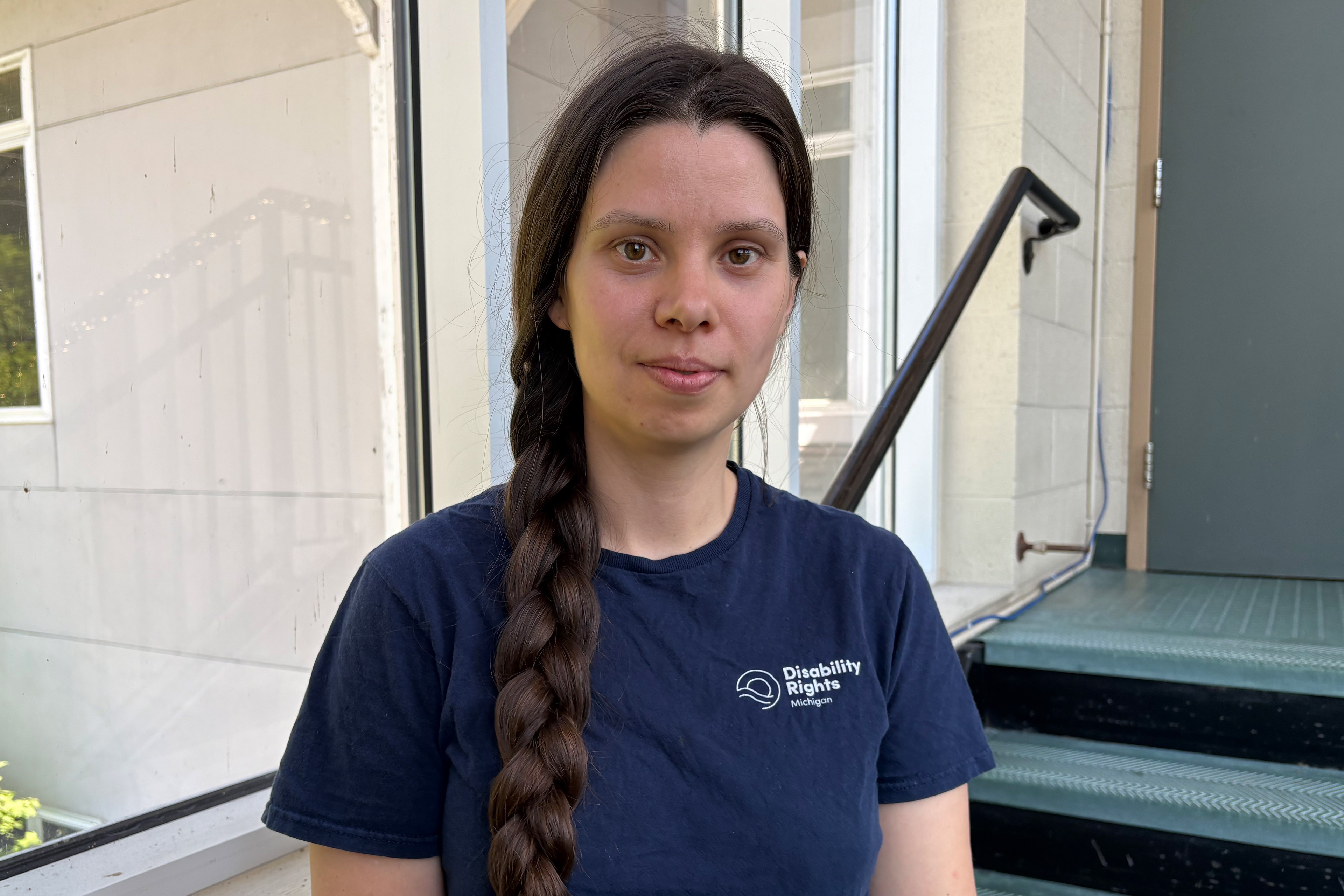
Anastassia Kolosova, a supervising attorney with Disability Rights Michigan, an organization dedicated to advocating for individuals with disabilities, has been unable to secure a meeting with Michigan officials to gain a deeper understanding of the specific system flaw that led to Noon’s coverage denial. This lack of transparency raises concerns about accountability and the state’s commitment to resolving systemic issues.
Deloitte’s long-standing involvement with Michigan’s Bridges system, which manages eligibility for Medicaid, SNAP, and other benefits, dates back at least 14 years. In its pursuit of continued contracts, the company frequently touts its extensive experience across the nation in Medicaid operations. Deloitte’s bid documents highlight their familiarity with the Bridges system, stating that their history in Michigan makes them "the ideal vendor" and that Michigan benefits from their "technical expertise drawn from across the nation" due to their work on similar systems in 31 other states.
However, advocates for individuals with disabilities contend that Michigan’s computer system has consistently failed to accurately recognize when certain adults should qualify for Medicaid benefits. This sentiment is echoed in other states where Deloitte operates. In Tennessee, a 2020 class-action lawsuit filed by Medicaid beneficiaries alleged that the state’s Deloitte-built system "does not reliably test for eligibility" for several categories of individuals with disabilities. The contract with Deloitte in Tennessee is reportedly valued at $1.12 billion over a decade. In 2024, a federal judge sided with the Medicaid beneficiaries, ruling that Tennessee had violated federal law and the U.S. Constitution. Notably, Deloitte was not named as a defendant in this lawsuit.
Further historical evidence of systemic issues in Michigan emerged from a 2010 report by the state’s Office of the Auditor General. This report found that government agencies had "did not provide effective project administration" and failed to ensure the state’s ability to "independently maintain and operate Bridges." The audit attributed this deficiency to the contractor’s failure to adequately transfer knowledge and skills to state officials. Consequently, Deloitte’s initial contract, valued at approximately $70 million, saw an increase of $50 million, a 71% escalation. State records indicate that subsequent additions brought the total cost of Deloitte’s initial contract to $124.1 million, with the auditor’s report forecasting "significant additional costs" for ongoing contract maintenance.
Despite these past findings, Sutfin asserts that "the state is now fully capable of operating and maintaining Bridges independently." Deloitte’s latest contract in Michigan, valued at $197.4 million, is set to expire in 2030.
A Pattern of Incorrect Coverage and Denials
Marie Noon’s application for Medicaid was submitted in August, and by September, she received a denial notice from the Michigan Department of Health and Human Services. The notice cited inaccurate income information and stated she was not disabled, according to Kolosova and state documents. Noon recounted her attempts to seek assistance from state workers, who "didn’t know anything about" the specific Medicaid program she had applied for, the "Freedom to Work" program. "I can’t tell you how many times I just wanted to give up," she admitted.
For individuals with disabilities participating in programs like "Freedom to Work," only half of their earnings are supposed to be considered when assessing Medicaid eligibility. This crucial provision was apparently not applied in Noon’s case. Kolosova believes that Michigan’s eligibility system failed to identify Noon as disabled, despite the state possessing all the necessary documentation to confirm her status. By misclassifying her disability, the state applied the incorrect income formula, leading to the erroneous conclusion that Noon earned too much to qualify for Medicaid. Both Deloitte and the Michigan Department of Health and Human Services declined to provide detailed responses to specific questions regarding Noon’s experience.
Kolosova noted a concerning increase in calls to Disability Rights Michigan from individuals experiencing denials related to the "Freedom to Work" benefit, averaging "two or three a month." She expressed concern that "There’s something wrong with the system if they’re relying on individual caseworkers to catch this. The system needs to work."
Noon’s situation is not an isolated incident. Similar errors have been reported in other states where Deloitte manages eligibility systems. In Texas, in 2023, Lilly Livingston, a 22-year-old with Down syndrome who requires extensive medical care including speech and occupational therapy, was abruptly removed from Medicaid. Her mother, Marie Livingston, reported that her daughter was mistakenly enrolled in "Healthy Texas Women," a program offering limited services primarily focused on breast and cervical cancer screenings and family planning. "Trying to fix that was a nightmare," Marie Livingston stated.
Terry Anstee, an attorney with Disability Rights Texas, intervened in Livingston’s case. In a September 2023 email flagged as "URGENT," Anstee pleaded with a Texas Medicaid eligibility worker for assistance, highlighting an unspecified "error" that had led to Livingston’s benefits being revoked. Anstee emphasized that Livingston’s recovery from two major surgeries was contingent on Medicaid coverage and that her eligibility through multiple pathways made the denial inexplicable. Deloitte declined to comment on Livingston’s case. Jennifer Ruffcorn, a spokesperson for Texas Health and Human Services, confirmed Livingston was erroneously enrolled in Healthy Texas Women but stated she did not experience a lapse in coverage in 2023. Anstee disputed this, asserting that Livingston did experience gaps in coverage during August and September 2023.
A similar issue was reported in Michigan in 2024, where advocates for mental health services raised concerns about individuals with disabilities being automatically enrolled in "Plan First," a limited Medicaid program focused on sexual health and family planning services. This program does not provide the comprehensive care required by individuals with disabilities. Malcolm Kletke, a lobbyist representing the Community Mental Health Association of Michigan and other mental health providers, wrote to a Michigan health official, expressing that enrollees who had long received Medicaid due to their disability were losing access to "services essential to their recovery and quality of life" by being incorrectly placed in Plan First.
State records supported these concerns, indicating a 10% decline in Medicaid enrollment within the category that includes individuals with disabilities over approximately four years, according to the Michigan House Fiscal Agency. This drop was considered unusual, as individuals typically leave the program due to death or recovery from temporary disabilities, making such a significant decrease unlikely without systemic issues. Robert Sheehan, former CEO of the mental health association, noted the anomaly.
In response to inquiries from KFF Health News, the Michigan health department acknowledged in April that changes had been made "to address concerns raised by advocates." Sutfin stated that the state’s computer system now prevents the approval of Plan First benefits until all other coverage options have been evaluated. She clarified that these changes were implemented "not to correct system errors," but rather to address the concerns raised. Sutfin further indicated that a change request was submitted to Deloitte to address this specific problem, with the fix implemented in January 2025. Prior to Kletke’s email, the state had reportedly denied the existence of problems related to Plan First.
Despite these adjustments, Noon’s struggle continued. Her coverage denial notice arrived in September, and she spent months appealing the decision, bearing the cost of her medications out-of-pocket throughout the appeals process. It was not until January that her Medicaid coverage was finally reinstated. "I literally cried," Noon said. "It was a really big deal."
The ongoing reliance on complex IT systems for essential safety net programs like Medicaid, particularly for vulnerable populations such as individuals with disabilities, highlights the critical need for robust oversight, transparent accountability, and systems that are designed with human needs at their core. The experiences of Marie Noon and others underscore the potentially devastating consequences when these systems falter, leaving individuals struggling to access the vital healthcare they depend on.